
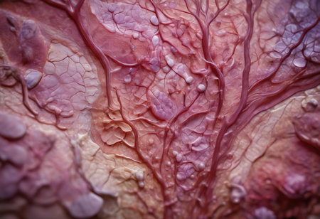
close up of red cabbage leaf with water droplets, abstract background
Коллекция по умолчанию
Коллекция по умолчанию
Создать новую
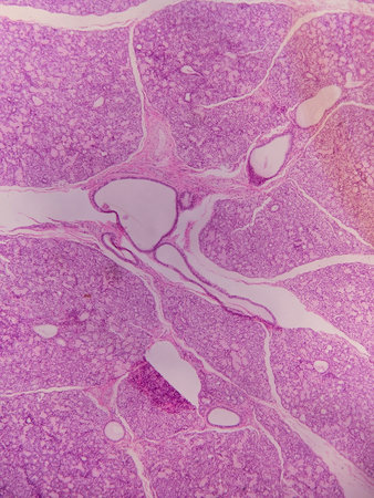
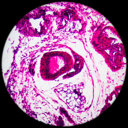
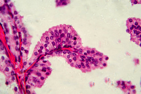
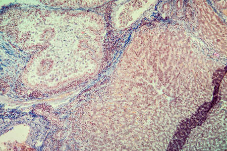
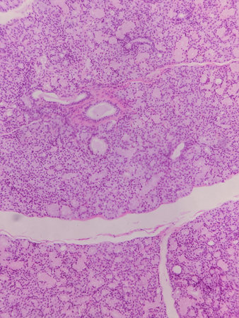
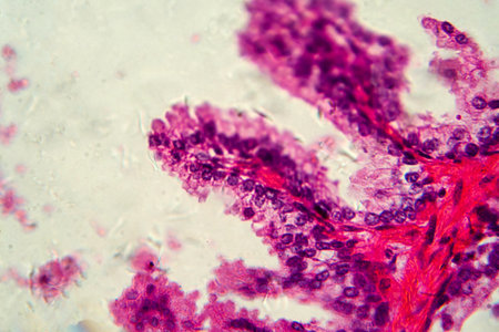
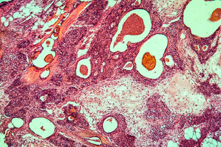
A microscopic view of tissue showing pink-stained cells and structures, indicative of biological samples, possibly related to histology or pathology
Коллекция по умолчанию
Коллекция по умолчанию
Создать новую
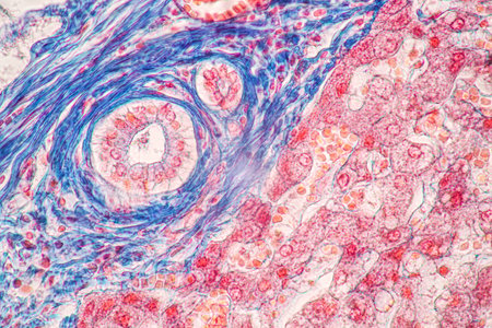
A close up of a plant organ with blue and red colors. The organ is surrounded by a white background
Коллекция по умолчанию
Коллекция по умолчанию
Создать новую
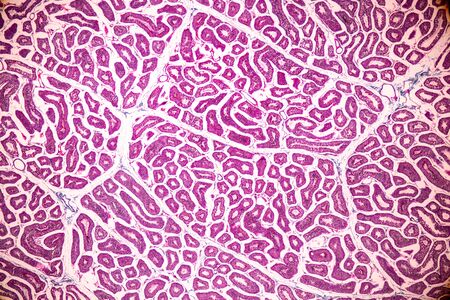
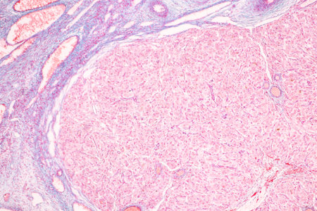
Structure of Tissue of Spleen Human, Liver Human and Kidney Human under the microscope in Lab.
Коллекция по умолчанию
Коллекция по умолчанию
Создать новую
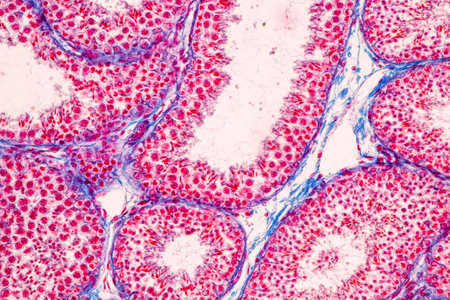
Anatomy and Histological Epididymis and Testis human cells under microscope.
Коллекция по умолчанию
Коллекция по умолчанию
Создать новую

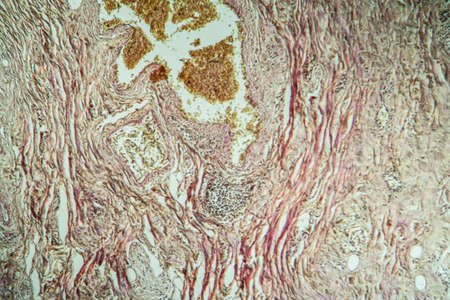
diseased liver with cirrhosis 100x under the microscope
Коллекция по умолчанию
Коллекция по умолчанию
Создать новую
Vibrant cross-section of a developing seed under UV light, highlighting the embryo and endosperm in bright colors
Коллекция по умолчанию
Коллекция по умолчанию
Создать новую
Anatomy and Histological Epididymis and Testis human cells under microscope.
Коллекция по умолчанию
Коллекция по умолчанию
Создать новую
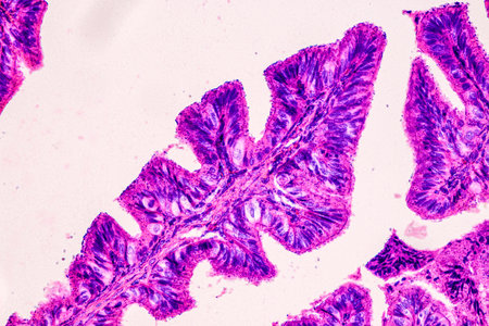
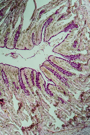
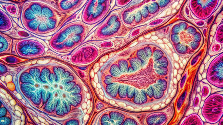
Tissue of Stomach Human under the microscope in Lab.
Коллекция по умолчанию
Коллекция по умолчанию
Создать новую
macro shot of liver tissue under a microscope, created with generative ai
Коллекция по умолчанию
Коллекция по умолчанию
Создать новую

Stomach tissue under the microscope 100x
Коллекция по умолчанию
Коллекция по умолчанию
Создать новую
Inguinal testicles gonadically diseased tissue 100x
Коллекция по умолчанию
Коллекция по умолчанию
Создать новую
Embryo with endosperm across 100x
Коллекция по умолчанию
Коллекция по умолчанию
Создать новую
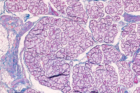
serous gland tissue under the microscope 100x
Коллекция по умолчанию
Коллекция по умолчанию
Создать новую
Anatomy and Histological Ovary, Testis and Sperm human cells under microscope.
Коллекция по умолчанию
Коллекция по умолчанию
Создать новую
Fresh Fruits Photography on Clean Background
Коллекция по умолчанию
Коллекция по умолчанию
Создать новую
A close up of a pink and blue cell with a tree inside of it. The cell is surrounded by a pink and blue background
Коллекция по умолчанию
Коллекция по умолчанию
Создать новую
Nestwurz orchid root cross 100x
Коллекция по умолчанию
Коллекция по умолчанию
Создать новую
science medical anthropotomy physiology microscopic section of lymph gland tissue background
Коллекция по умолчанию
Коллекция по умолчанию
Создать новую
3D illustration of the human organ systems, Human internal organs. Anatomy. Nervous, circulatory, digestive, excretory, urinary,and bone systems. Medical education concept, Generative AI illustration
Коллекция по умолчанию
Коллекция по умолчанию
Создать новую

Thyroid follicular carcinoma, light micrograph, photo under microscope
Коллекция по умолчанию
Коллекция по умолчанию
Создать новую
Microscopic view of human pancreas in laboratory. 3D rendering
Коллекция по умолчанию
Коллекция по умолчанию
Создать новую
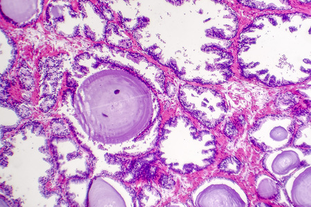
Goiter colloid goiter disease 100x
Коллекция по умолчанию
Коллекция по умолчанию
Создать новую
Education anatomy and Histological sample of Human under the microscope.
Коллекция по умолчанию
Коллекция по умолчанию
Создать новую
Abstract marbling floral pattern for fabric, tile design. background texture
Коллекция по умолчанию
Коллекция по умолчанию
Создать новую
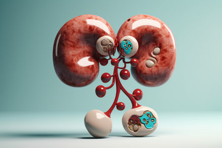
Kidneys. Cute cartoon healthy human anatomy internal organ character set with brain lung intestine heart kidney liver and stomach mascots. parts of living body organs in animated form
Коллекция по умолчанию
Коллекция по умолчанию
Создать новую
This detailed microscopic image showcases various cellular structures, highlighted in striking purple tones. The intricate patterns and textures reveal the complexity of biological tissues, making it a valuable resource for educational and scientific purposes
Коллекция по умолчанию
Коллекция по умолчанию
Создать новую
Breast tissue under the microscope 100x
Коллекция по умолчанию
Коллекция по умолчанию
Создать новую

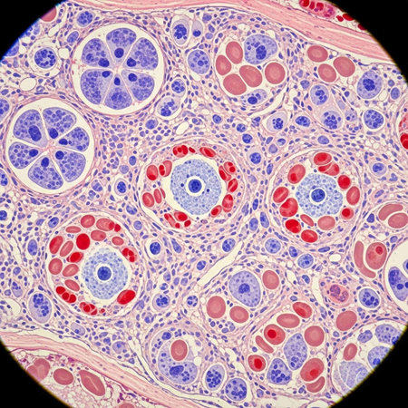
Lacrimal gland tissue under the microscope 100x
Коллекция по умолчанию
Коллекция по умолчанию
Создать новую
Columnar epithelium of human gall bladder under the microscope in Lab.
Коллекция по умолчанию
Коллекция по умолчанию
Создать новую
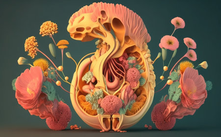
Human anatomy, 3D illustration. Human digestive system with flowers and plants.
Коллекция по умолчанию
Коллекция по умолчанию
Создать новую
Histopathology of cirrhosis, light micrograph, photo under microscope
Коллекция по умолчанию
Коллекция по умолчанию
Создать новую
science medical anthropotomy physiology microscopic section of human thyroid gland background
Коллекция по умолчанию
Коллекция по умолчанию
Создать новую

Anatomy and Histological Epididymis and Testis human cells under microscope.
Коллекция по умолчанию
Коллекция по умолчанию
Создать новую
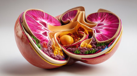
A close-up of the human kidney with visible structures such as nephrons and blood vessels, set on a neutral background with a focus on details
Коллекция по умолчанию
Коллекция по умолчанию
Создать новую
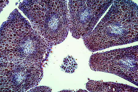
Palatal tonsils transverse 100x under a microscope
Коллекция по умолчанию
Коллекция по умолчанию
Создать новую
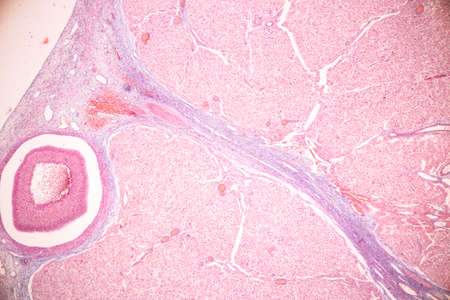
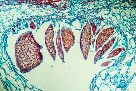
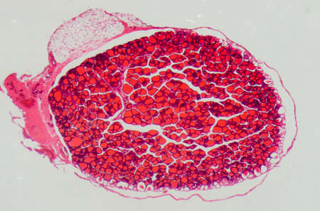
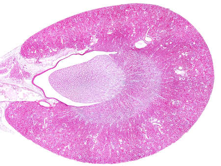
Cross section Human testis under microscope view. Shows spermatogonia, spermatocytes in meiosis, spermatids, and spermatozoa
Коллекция по умолчанию
Коллекция по умолчанию
Создать новую
Cross section of Testis tissue under the microscope for education.
Коллекция по умолчанию
Коллекция по умолчанию
Создать новую
destructive mushroom in wood fabric 100x
Коллекция по умолчанию
Коллекция по умолчанию
Создать новую

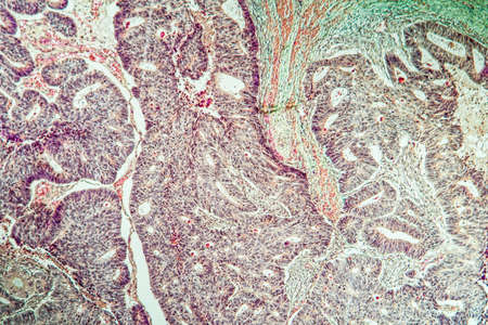
Ovarian cancer, light micrograph, photo under microscope. Photograph shows a fragment of a cancerous tumor in the female ovary. Selective focus
Коллекция по умолчанию
Коллекция по умолчанию
Создать новую
Uterus and ovaries. Women health symbol. Female reproductive and sexual health. Reproductive system model uterus between two palms. Female reproductive organs. Questions of conception and gestation.
Коллекция по умолчанию
Коллекция по умолчанию
Создать новую
Leech cross section showing internal anatomical structures stained
Коллекция по умолчанию
Коллекция по умолчанию
Создать новую

Bladder cancer, light micrograph, photo under microscope
Коллекция по умолчанию
Коллекция по умолчанию
Создать новую
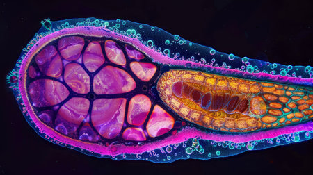
Vibrant cross-section of a developing seed under UV light, highlighting the embryo and endosperm in bright colors
Коллекция по умолчанию
Коллекция по умолчанию
Создать новую
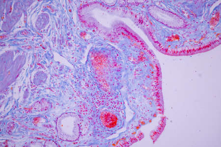
Photomicrograph showing histological features of benign prostatic hyperplasia. Enlarged prostate gland with nodular proliferation of glandular and stromal components. High-resolution histology image.
Коллекция по умолчанию
Коллекция по умолчанию
Создать новую
Colorful organic shapes flow and merge in a captivating abstract illustration, showcasing soft pink and orange tones set against a neutral backdrop, creating a serene visual experience.
Коллекция по умолчанию
Коллекция по умолчанию
Создать новую

Brain cells in the dark field under the microscope 100x
Коллекция по умолчанию
Коллекция по умолчанию
Создать новую

Histological Spermatic cord human, Seminal vesicle human, Prostate human and Human chromosomes under the microscope for education.
Коллекция по умолчанию
Коллекция по умолчанию
Создать новую
Education anatomy and Histological sample of Human under the microscope.
Коллекция по умолчанию
Коллекция по умолчанию
Создать новую
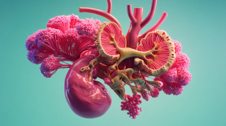
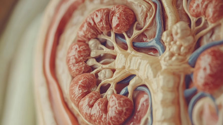
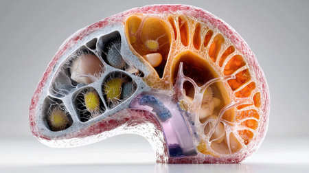
This vibrant 3D rendering showcases a human kidney with intricate vascular structures, ideal for educational and medical use. The creative design highlights the organ's anatomy and surrounding features in a visually appealing format.
Коллекция по умолчанию
Коллекция по умолчанию
Создать новую
Coccidiosis of liver tissue under the microscope 100x
Коллекция по умолчанию
Коллекция по умолчанию
Создать новую
A stunning cross-sectional illustration of a fruit revealing vibrant colors and intricate internal structures, perfect for educational and artistic purposes.
Коллекция по умолчанию
Коллекция по умолчанию
Создать новую
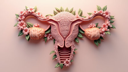
Organ of the uterus with flowers, female nature. Woman reproductive health illustration.
Коллекция по умолчанию
Коллекция по умолчанию
Создать новую
Abstract representation of biological cells with interconnected structures, showcasing vibrant red and blue tones. Conceptual depiction of cellular activity and science.
Коллекция по умолчанию
Коллекция по умолчанию
Создать новую

Abstract hand drawn watercolor drop, for backgrounds or textures
Коллекция по умолчанию
Коллекция по умолчанию
Создать новую
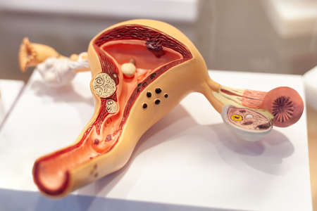
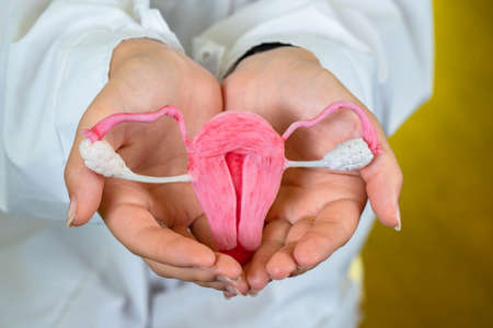
Model of sick uterus with ovaries in woman
Коллекция по умолчанию
Коллекция по умолчанию
Создать новую

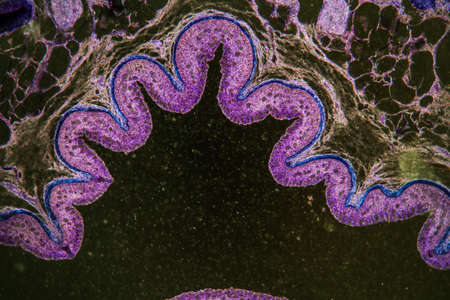
Characteristics Tissue of Olfactory epithelium Human under the microscope in Lab.
Коллекция по умолчанию
Коллекция по умолчанию
Создать новую
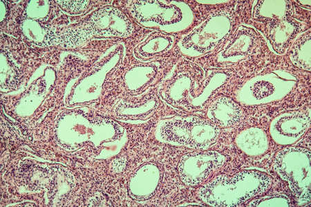
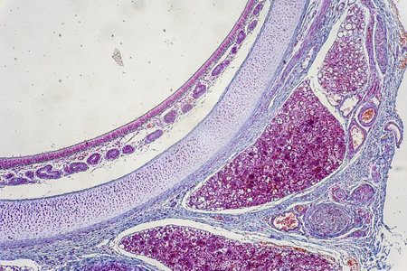
The study of tissue samples of Trachea of Cat, Epididymis, Prostate, Uterus with embryo of rat and Mammary gland cow under the microscope in Lab.
Коллекция по умолчанию
Коллекция по умолчанию
Создать новую

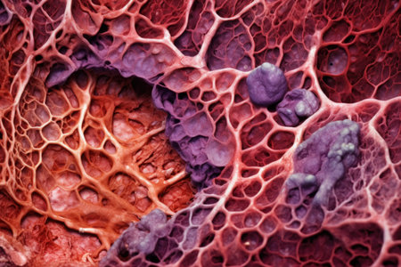
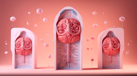
A cancer cell in a person with abnormal cells. A cancer cell on an internal organ. The concept of medical science.
Коллекция по умолчанию
Коллекция по умолчанию
Создать новую

Anatomy and Histological Epididymis and Testis human cells under microscope.
Коллекция по умолчанию
Коллекция по умолчанию
Создать новую
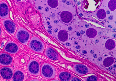
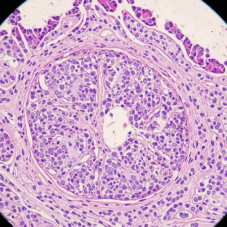
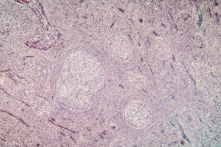
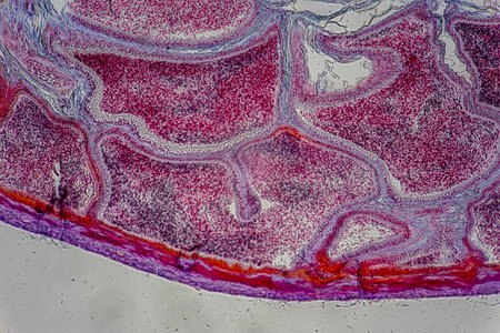
Showing Light micrograph of the Thyroid gland and Thymus gland human Child under the microscope for education in the laboratory.
Коллекция по умолчанию
Коллекция по умолчанию
Создать новую
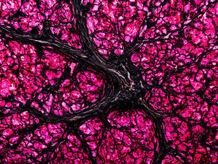
Histopathology of human liver under microscope view. Histological sample of human liver.
Коллекция по умолчанию
Коллекция по умолчанию
Создать новую
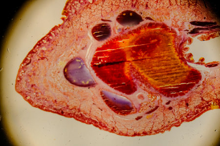
Fish anatomy. Pike (Esox lucius). The hepatic parenchyma is divided into lobules and covered with a fibrous membrane. Digestive, metabolic and protective function. Extreme close up
Коллекция по умолчанию
Коллекция по умолчанию
Создать новую
Anatomy and Histological Ovary, Testis and Sperm human cells under microscope.
Коллекция по умолчанию
Коллекция по умолчанию
Создать новую
The study of tissue samples of Trachea of Cat, Epididymis, Prostate, Uterus with embryo of rat and Mammary gland cow under the microscope in Lab.
Коллекция по умолчанию
Коллекция по умолчанию
Создать новую
Photomicrograph showing histological features of benign prostatic hyperplasia. Enlarged prostate gland with nodular proliferation of glandular and stromal components.
Коллекция по умолчанию
Коллекция по умолчанию
Создать новую

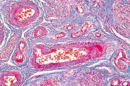
Anatomy and Histological Bone, Elastic cartilage human and Joint of human foetus under the microscope for education.
Коллекция по умолчанию
Коллекция по умолчанию
Создать новую

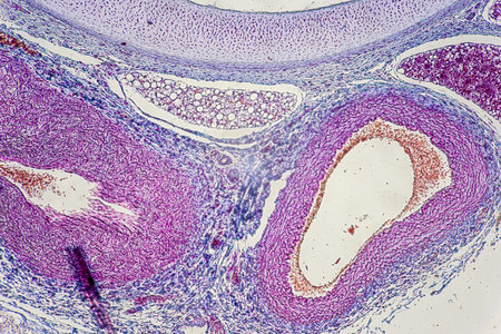
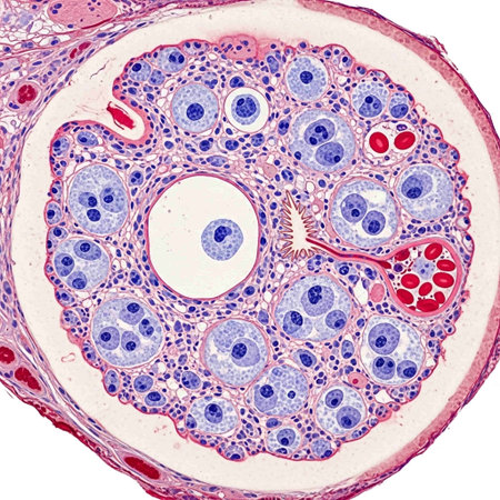
Light micrograph of the Cat's ovarian. Micrograph of ovary showing primordial, primary and secondary follicles isolated on white background. Hematoxylin and eosin stain.
Коллекция по умолчанию
Коллекция по умолчанию
Создать новую
Generative AI Closeup shot of loofah body scrub
Коллекция по умолчанию
Коллекция по умолчанию
Создать новую
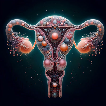
Digital illustration of a female reproductive system in colored background with copy space
Коллекция по умолчанию
Коллекция по умолчанию
Создать новую
Colon carcinoma arising from adenoma, 100x
Коллекция по умолчанию
Коллекция по умолчанию
Создать новую
Photomicrograph showing histological features of benign prostatic hyperplasia. Enlarged prostate gland with nodular proliferation of glandular and stromal components. High-resolution histology image.
Коллекция по умолчанию
Коллекция по умолчанию
Создать новую
Anatomy and Histological Bone, Elastic cartilage human and Joint of human foetus under the microscope for education.
Коллекция по умолчанию
Коллекция по умолчанию
Создать новую
Histopathology of alveoli, light micrograph, photo under microscope
Коллекция по умолчанию
Коллекция по умолчанию
Создать новую
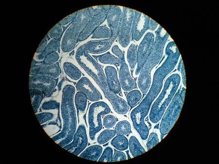
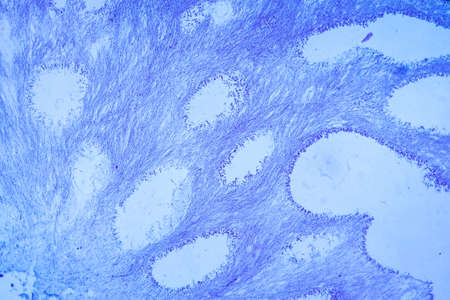
Cross-section through the intestine with glands 200x
Коллекция по умолчанию
Коллекция по умолчанию
Создать новую
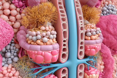
This image depicts a magnified cross-section of the human pancreas, revealing its intricate structure with visible islets of Langerhans, acinar cells, and pancreatic ducts.
Коллекция по умолчанию
Коллекция по умолчанию
Создать новую
Breast tissue under the microscope 100x
Коллекция по умолчанию
Коллекция по умолчанию
Создать новую
Colon tissue with diverticulum 100x
Коллекция по умолчанию
Коллекция по умолчанию
Создать новую
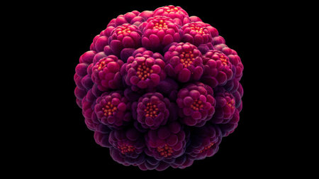
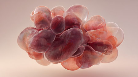
This abstract image features a spherical cluster in vibrant pink and purple against a dark backdrop, highlighting intricate organic shapes and artistic design.
Коллекция по умолчанию
Коллекция по умолчанию
Создать новую
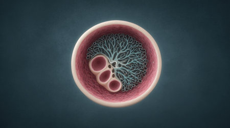
This visual showcases a comprehensive 3D cross-section of nephron anatomy with clean zones, displaying intricate cellular structures and arrangements.
Коллекция по умолчанию
Коллекция по умолчанию
Создать новую
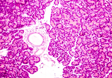
Microscopic photo showing pancreatic tissue. Light micrograph of pancreas, magnification 100x
Коллекция по умолчанию
Коллекция по умолчанию
Создать новую
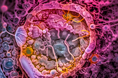
Microscopic macro close up shot of human cell. Biology, microbiology or chemistry as scientific abstract background. Human cell or molecule virus cell structure visualization. generative ai
Коллекция по умолчанию
Коллекция по умолчанию
Создать новую
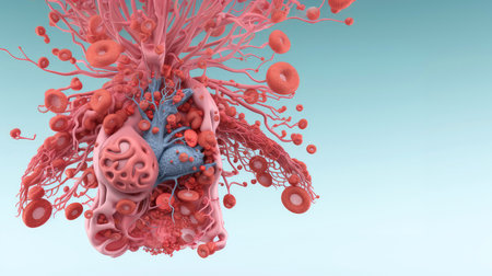
A close up of a human organ with a pinkish color. The organ is a heart and it is shown in a very detailed manner. Concept of awe and wonder at the complexity of the human body
Коллекция по умолчанию
Коллекция по умолчанию
Создать новую
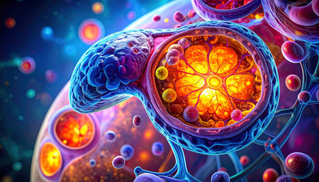
Detailed cross-section of a vibrant purple cell showing glowing orange internal structures and surrounding...
Коллекция по умолчанию
Коллекция по умолчанию
Создать новую
Cancer swollen Parotid diseased tissue 100x
Коллекция по умолчанию
Коллекция по умолчанию
Создать новую
Bowel with goblet cells in the dark field 100x
Коллекция по умолчанию
Коллекция по умолчанию
Создать новую
Histopathology of fibroids, light micrograph, photo under microscope
Коллекция по умолчанию
Коллекция по умолчанию
Создать новую

Histological Spermatic cord human, Seminal vesicle human, Prostate human and Human chromosomes under the microscope for education.
Коллекция по умолчанию
Коллекция по умолчанию
Создать новую
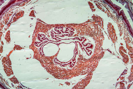
Light micrograph cross section of rat kidney stained with hematoxylin and eosin displaying the peripheral renal cortex (with many glomeruli) and, in the central medulla, a Malpighian pyramid leading to a renal calyx.
Коллекция по умолчанию
Коллекция по умолчанию
Создать новую
Rhodonite A deep pink rhodonite with black manganese oxide veins suspended and rotating to show
Коллекция по умолчанию
Коллекция по умолчанию
Создать новую
The study of tissue samples of Trachea of Cat, Epididymis, Prostate, Uterus with embryo of rat and Mammary gland cow under the microscope in Lab.
Коллекция по умолчанию
Коллекция по умолчанию
Создать новую

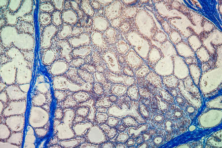
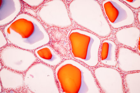
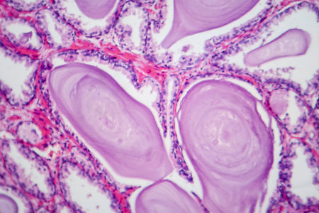
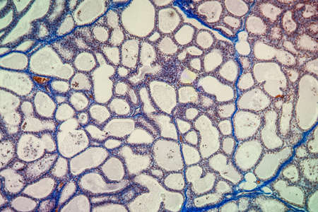
Low magnification micrograph showing a human thyroid. The thyroid parenchyma is composed by many follicle filled with colloid. Light microscope micrograph. Hematoxylin & eosin stain
Коллекция по умолчанию
Коллекция по умолчанию
Создать новую
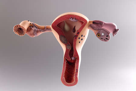
Model of the female reproductive system on gray background
Коллекция по умолчанию
Коллекция по умолчанию
Создать новую
Benign prostatic hyperplasia. Micrograph shows dilated glands, papillary projections inside the lumen of the glands, cystic dilatation with accumulation of secretory material. Photo under microscope
Коллекция по умолчанию
Коллекция по умолчанию
Создать новую
Spatter drop. Sparkling splash. Defocused purple red neon ink color rainbow round shiny texture spreading on abstract light creative background.
Коллекция по умолчанию
Коллекция по умолчанию
Создать новую
macro photo of a red skin texture
Коллекция по умолчанию
Коллекция по умолчанию
Создать новую
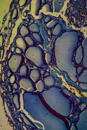
Close-up of plant stem cells under microscope, intricate patterns, high detail, with copy space
Коллекция по умолчанию
Коллекция по умолчанию
Создать новую
3D illustration of the human organ systems, Human internal organs. Anatomy. Nervous, circulatory, digestive, excretory, urinary,and bone systems. Medical education concept, Generative AI illustration
Коллекция по умолчанию
Коллекция по умолчанию
Создать новую
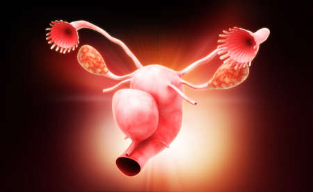
Digital illustration of female reproductive system in colour background
Коллекция по умолчанию
Коллекция по умолчанию
Создать новую

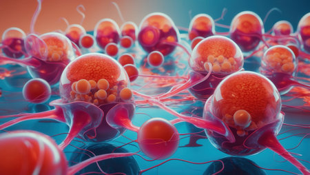
Cancer cells, Tumor growing in human body
Коллекция по умолчанию
Коллекция по умолчанию
Создать новую
Legion-Media
Создайте свои проекты на основе качественных стоковых фотографий и видео.


Copyright © Legion-Media.